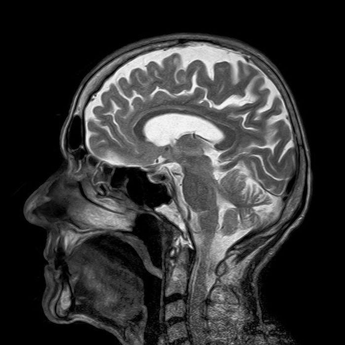
A Revolutionary Look Inside the Human Brain's Structures
Written on
Chapter 1: The Future of Brain Imaging
Envision a world where we can visualize the human brain in exceptional detail, allowing for the early detection of tumors or neurodegenerative conditions. Although we are not quite there yet, advancements in 3-dimensional (3D) imaging techniques are bringing us closer to this reality, offering a new perspective on the brain's anatomical features with remarkable clarity.
The examination of organ systems in 3D has been a staple in biomedical research for over a century, linking traditional anatomical knowledge with modern systems biology. Optical imaging methods frequently utilize fluorescent tags, which illuminate internal structures at a microscopic level. However, a significant challenge arises from the opacity of biological tissues, which can obscure fluorescent signals and compromise image quality. Recent innovations in tissue-clearing chemistry are addressing this issue, allowing researchers to unveil the previously concealed complexities of anatomical architecture.
Section 1.1: Breakthroughs at RIKEN Center
Innovative imaging scientists at the RIKEN Center for Biosystems Dynamics Research have introduced a groundbreaking method for visualizing internal structures, ranging from individual organs to entire organisms. By submerging biological samples in a specialized tissue-clearing gel and then treating them with a blend of tissue dyes and fluorescently-labeled antibodies, researchers can pinpoint specific cell populations and intricate tissue arrangements within organs. Their protocol was validated using brains from mice and marmosets, demonstrating its effectiveness.
Researchers published their findings in Nature Communications, highlighting the extraordinary capabilities of their novel imaging technique. The 3D renderings of scanned images from the brains of both species unveiled remarkable similarities in their neural vasculature. Furthermore, the team showcased an innovative approach to simultaneously label multiple molecular targets within the mouse brain.
Section 1.2: A Glimpse Inside the Mind
The technique was also applied to image a complete organism, specifically an infant marmoset, as well as a small sample of human brain tissue. This versatility suggests significant potential for the method to evolve into a valuable research or even diagnostic tool in the future.

Chapter 2: The Impact of Innovative Imaging Techniques
The first video offers a glimpse into the intriguing narrative of Charles Swan III, showcasing the complexities of the human mind and the experiences that shape it.
The second video features a clip from A Glimpse Inside the Mind of Charles Swan III, highlighting the artistic representation of psychological themes and emotional depth.